Double-outlet right ventricle
Overview
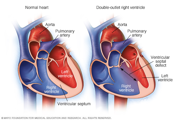
In double-outlet right ventricle, the aorta and pulmonary artery connect partially or completely to the right ventricle. A hole also exists between the two ventricles (ventricular septal defect). In a normal heart, as shown on the left, the pulmonary artery connects to the right ventricle and the aorta connects to the left ventricle.
Double-outlet right ventricle is a heart condition present at birth (congenital) in which two large blood vessels don't connect to the heart normally. In infants born with this condition, the main artery that carries blood from the heart to the body (aorta) and the artery that directs blood from the heart to the lungs (pulmonary artery) connect partially or completely to the right lower heart chamber (ventricle). Sometimes these blood vessels are also reversed from their normal positions.
In a normal heart, the aorta connects to the left ventricle and the pulmonary artery connects to the right ventricle.
In infants with double-outlet right ventricle, there's also a hole between the right and left ventricles (ventricular septal defect). This causes oxygen-rich blood to mix with oxygen-poor blood. Children born with double-outlet right ventricle can have a lower than normal amount of oxygen in the bloodstream.
If too much blood flows through the pulmonary artery to the lungs, it can cause heart failure and poor growth over time. In other cases, blood flow through the pulmonary artery may be reduced, which can cause your child's skin to turn a bluish color.
Some children will need surgery within the first few days of birth to correct the heart defect. Others may have surgery at a few months of age.
Also, some people with double-outlet right ventricle may have other associated congenital heart defects, including other holes in the heart, heart valve problems or blood vessel problems.
Diagnosis
Doctors will generally perform an echocardiogram to diagnose double-outlet right ventricle and any associated defects. Echocardiograms use sound waves to produce an image of the heart. Sound waves bounce off the heart and produce moving images that can be viewed on a video screen.
If more information is needed, doctors may conduct cardiac CT and MRI scans or cardiac catheterization. In cardiac catheterization, your child's doctor inserts a thin, flexible tube (catheter) into an artery or vein in the groin or neck and threads it into the heart. A dye is injected through the catheter to make the heart structures more visible on X-ray pictures. Cardiac catheterization also measures pressure and oxygen levels in the chambers of the heart and in the blood vessels.
Treatment
Several types of surgery may be performed, depending on the specific heart defect and any associated defects.
Depending on the type of defect, surgeons may:
- Create a tunnel through the ventricular septal defect to connect the left ventricle to the aorta
- Switch the aorta and pulmonary artery positions, if they're reversed, in order to connect the pulmonary artery to the right ventricle and the aorta to the left ventricle
- Insert a patch to close the hole between the ventricles
- Insert a blood vessel to connect the right ventricle to the pulmonary artery, allowing more blood flow if the pulmonary artery is small
- Widen a narrowed pulmonary artery to allow more blood flow
- Conduct a series of other procedures to allow blood to move to the lungs and for the heart to function with one ventricle, if the defect is complex
- Repair any other congenital heart defects
In some infants with inadequate blood flow to their lungs at birth, a temporary procedure may be done to insert a shunt between the aorta and the pulmonary artery. The shunt is removed later in life during heart surgery to repair the defect.
Adults who were born with double-outlet right ventricle will need lifelong care and regular follow-up exams. They should see a heart doctor trained in evaluating and treating congenital heart conditions (adult congenital cardiologist).
Surgery may be needed later in life for valve disease if there's a narrowing or leakage of the heart valves. Some adults need close monitoring of their aortas and pulmonary arteries, especially if they required surgery early in life. A small group of adults may require medications for treatment of decreased function involving the right or left ventricles.
Content Last Updated: January 6, 2021
Content provided by Mayo Clinic ©1998-2026 Mayo Foundation for Medical Education and Research (MFMER). All rights reserved. Terms of Use