Brain stereotactic radiosurgery
Overview
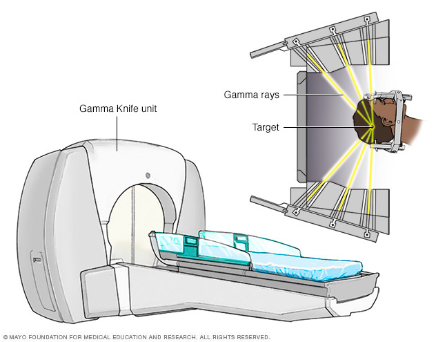
Gamma Knife stereotactic radiosurgery technology uses many small gamma rays to deliver a precise dose of radiation to a target.
Gamma Knife radiosurgery is a type of radiation therapy used to treat tumors, vascular malformations and other abnormalities in the brain.
Gamma Knife radiosurgery, like other forms of stereotactic radiosurgery (SRS), is not surgery in the traditional sense because there is no incision.
Instead, Gamma Knife radiosurgery uses specialized equipment to focus about 200 tiny beams of radiation on a tumor or other target with submillimeter accuracy. Although each beam has very little effect on the brain tissue it passes through, a strong dose of radiation is delivered to the place where all the beams meet.
The precision of brain stereotactic radiosurgery results in minimal radiation delivery to healthy tissues surrounding the target.
Gamma Knife radiosurgery is usually a one-time therapy completed in a single day.
Why it’s done
Gamma Knife radiosurgery is often a safer alternative to standard brain surgery (neurosurgery), which requires incisions in the scalp, an opening in the skull and membranes surrounding the brain, and dissection into brain tissue. This type of radiation treatment is usually performed when:
- A tumor or other abnormality in the brain is too hard to reach with standard neurosurgery
- A person isn’t healthy enough to undergo standard surgery
- A person prefers a less invasive treatment
In some cases, Gamma Knife radiosurgery may have a lower risk of side effects compared with other types of radiation therapy and it can all be done in one day compared with up to 30 treatments with conventional radiation therapy.
Gamma Knife radiosurgery is most commonly used to treat the following conditions:
-
Brain tumor. Radiosurgery is useful in the management of small noncancerous (benign) and cancerous (malignant) brain tumors.
Radiosurgery damages the genetic material (DNA) in the tumor cells. The cells lose their ability to reproduce and may die, and the tumor may gradually shrink.
-
Arteriovenous malformation (AVM). AVMs are abnormal tangles of arteries and veins in your brain. In an AVM, blood flows from your arteries to veins, bypassing smaller blood vessels (capillaries). AVMs, if left untreated, may “steal” the normal flow of blood from the brain, causing a stroke, or lead to bleeding in the brain.
Radiosurgery destroys the AVM and causes the blood vessels to close off over time.
-
Trigeminal neuralgia. Trigeminal neuralgia is a disorder of one or both of the trigeminal nerves, which relay sensory information between your brain and areas of your forehead, cheek and lower jaw. This nerve disorder causes disabling facial pain that feels like an electric shock.
After treatment, many people will experience pain relief within a few days to a few months.
-
Acoustic neuroma. An acoustic neuroma (vestibular schwannoma) is a noncancerous (benign) tumor that develops along the nerve of balance and hearing leading from your inner ear to your brain.
When the tumor puts pressure on the nerve, you can experience hearing loss, dizziness, loss of balance and ringing in the ear (tinnitus). As the tumor grows, it can also put pressure on the nerves affecting sensations and muscle movement in the face.
Radiosurgery may stop the growth of an acoustic neuroma.
-
Pituitary tumors. Tumors of the bean-sized gland at the base of the brain (pituitary gland) can cause a variety of problems. The pituitary gland regulates hormones in your body that control various functions, such as your stress response, metabolism and sexual function.
Radiosurgery can be used to shrink the tumor and lessen the disruption of pituitary hormone regulation.
Risks
Gamma Knife radiosurgery doesn’t involve surgical incisions, so it’s generally less risky than traditional neurosurgery. In traditional neurosurgery, there are potential complications associated with anesthesia, bleeding and infection.
Early complications or side effects are usually temporary. Some people experience mild headaches, a tingling sensation on the scalp, nausea or vomiting. Other side effects may include:
- Fatigue. Tiredness and fatigue may occur for the first few weeks after Gamma Knife radiosurgery.
- Swelling. Swelling in the brain at or near the treatment site can cause a variety of symptoms depending on what areas of the brain are involved. If post-treatment swelling and symptoms do occur from the Gamma Knife treatment, these symptoms usually show up approximately six months after treatment rather than immediately after the procedure like with conventional surgery. Your doctor may prescribe anti-inflammatory medications (corticosteroid medications) to prevent such problems or to treat symptoms if they appear.
-
Scalp and hair problems. Your scalp may be red, irritated or sensitive at the four sites where the head frame was attached to your head during the treatment. But the head frame does not leave any permanent marks on the scalp. Rarely, some people temporarily lose a small amount of hair if the area being treated is right under the scalp.
Rarely, people may experience late side effects, such as other brain or neurological problems, months after Gamma Knife radiosurgery.
How you prepare
Food and medications
- Don’t eat or drink anything after midnight the night before the procedure.
- Talk to your doctor about whether you can take your regular medications the night before or morning of the procedure.
Clothing and personal items
Wear comfortable, loosefitting clothing.
Avoid wearing the following items during the procedure:
- Jewelry
- Eyeglasses
- Contact lenses
- Makeup
- Nail polish
- Dentures
- Wigs or hairpieces
Other precautions
Tell your doctor if you:
- Are taking pills or injections to control diabetes
- Are allergic to shellfish or iodine — both are chemically related to special dyes that may be used during the procedure
- Have implanted medical devices in your body, such as a pacemaker, artificial heart valve, aneurysm clips, neurostimulators or stents
- Experience claustrophobia
What you can expect
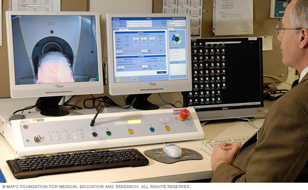
A radiation oncologist monitors the Gamma Knife procedure in progress.
Before the procedure
Before the procedure begins, you’ll have a lightweight frame attached to your head with four pins. This frame will stabilize your head during the radiation treatment and serve as a point of reference for focusing the beams of radiation. During this process:
- Your hair will not be shaved, but your hair may be washed with a special shampoo
- You’ll receive numbing shots in the four places on your scalp where the pins will be inserted — two points on your forehead and two at the back of your head
After the head frame is attached, you’ll undergo imaging scans of your brain that show the location of the tumor or other abnormality in relation to the head frame. The type of scan used depends on the condition being treated:
-
Tumors. Imaging for tumors may include computerized tomography (CT) or magnetic resonance imaging (MRI). In a CT scan, a series of X-rays creates a detailed image of your brain. In an MRI scan, a magnetic field and radio waves create detailed images of your brain.
A small needle may be placed in the back of your hand or in your arm to inject a dye into a blood vessel to view the blood vessels in your brain and highlight blood circulation. In some cases, you may have both MRI and CT scans.
-
Arteriovenous malformations (AVMs). Imaging for brain AVMs may include CT scans, MRI scans, cerebral angiograms or some combination of these tests.
In a cerebral angiogram, a doctor inserts a small tube in a blood vessel in your groin and threads it to the brain using X-ray imaging. Dye is injected through the blood vessels to make them visible on X-rays. Your doctor may inject a dye into a blood vessel during CT or MRI scans to view the blood vessels and highlight blood circulation.
- Trigeminal neuralgia. An MRI or a CT scan is used to create images of nerve fibers to select a target area for treating trigeminal neuralgia.
The results of the brain scans are fed into a computerized planning system that allows the radiosurgery team to determine the appropriate areas to treat, doses of radiation and how to focus the radiation beams to treat the areas. This planning process may take an hour or two. During that time, you can relax in another room, but the frame must remain attached to your head.
Children are often anesthetized for the imaging tests and during the radiosurgery. Adults are usually awake, but may be given a mild sedative to help them relax.
During the procedure
You’ll lie on a bed that slides into the Gamma Knife machine, and your head frame will be attached securely to a helmet inside the machine.
You’ll have an intravenous (IV) tube that delivers fluids to your bloodstream to keep you hydrated during the day. A needle at the end of the IV is placed in a vein, most likely in your arm.
The time needed to complete the procedure may range from less than an hour to about four hours, depending on the size and shape of the target. During the procedure:
- You won’t feel the radiation
- You won’t hear any noise from the machine
- You’ll be able to talk with the doctors via a microphone
Gamma Knife radiosurgery is usually an outpatient procedure, but the entire process will take most of a day. You may be advised to have a family member or friend who can be with you during the day and who can take you home. In some cases, an overnight stay in the hospital may be necessary.
After the procedure
After the procedure, you can expect the following:
- The head frame will be removed.
- You may have minor bleeding or tenderness at the pin sites.
- If you experience headache, nausea or vomiting after the procedure, you’ll receive appropriate medications.
- You’ll be able to eat and drink after the procedure.
Results
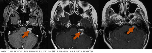
Gamma Knife radiosurgery uses multiple, tiny beams of radiation to shrink tumors. The left brain scan shows a pretreatment image of a noncancerous tumor — an image enhanced by the use of a special dye (contrast agent). At six months after treatment (middle image), the tumor appears slightly larger but doesn’t take up as much of the contrast agent — isn’t as bright in the center — indicating a positive treatment effect. At seven years (right image), the tumor appears much smaller.
The treatment effect of Gamma Knife radiosurgery occurs gradually, depending on the condition being treated:
- Benign tumors. Gamma Knife radiosurgery results in the failure of tumor cells to reproduce. The tumor may shrink over a period of 18 months to two years, but the main goal of Gamma Knife radiosurgery for benign tumors is to prevent any future tumor growth.
- Malignant tumors. Cancerous (malignant) tumors may shrink more rapidly, often within a few months.
- Arteriovenous malformations (AVMs). The radiation therapy causes the abnormal blood vessels of brain AVMs to thicken and close off. This process may take two years or more.
-
Trigeminal neuralgia. Gamma Knife radiosurgery creates a lesion that blocks transmission of pain signals along the trigeminal nerve. Pain relief may take several months.
You’ll receive instruction on appropriate follow-up exams to monitor your progress.
Content Last Updated: May 13, 2021
Content provided by Mayo Clinic ©1998-2022 Mayo Foundation for Medical Education and Research (MFMER). All rights reserved. Terms of Use