Brain AVM (arteriovenous malformation)
Overview
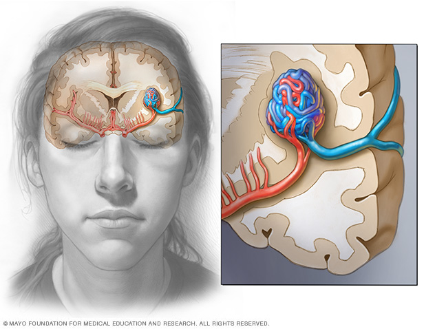
In a brain AVM, blood passes directly from your arteries to your veins via abnormal vessels. This disrupts the normal process of how blood circulates through your brain.
A brain arteriovenous malformation (AVM) is a tangle of abnormal blood vessels connecting arteries and veins in the brain.
The arteries are responsible for taking oxygen-rich blood from the heart to the brain. Veins carry the oxygen-depleted blood back to the lungs and heart. A brain AVM disrupts this vital process.
An arteriovenous malformation can develop anywhere in your body but occurs most often in the brain or spine. Even so, brain AVMs are rare and affect less than 1 percent of the population.
The cause of AVMs is not clear. Most people are born with them, but they can occasionally form later in life. They are rarely passed down among families genetically.
Some people with brain AVMs experience signs and symptoms, such as headache or seizures. AVMs are commonly found after a brain scan for another health issue or after the blood vessels rupture and cause bleeding in the brain (hemorrhage).
Once diagnosed, a brain AVM can often be treated successfully to prevent complications, such as brain damage or stroke.
Symptoms
A brain arteriovenous malformation may not cause any signs or symptoms until the AVM ruptures, resulting in bleeding in the brain (hemorrhage). In about half of all brain AVMs, hemorrhage is the first sign.
But some people with brain AVM may experience signs and symptoms other than bleeding related to the AVM.
In people without hemorrhage, signs and symptoms of a brain AVM may include:
- Seizures
- Headache or pain in one area of the head
- Muscle weakness or numbness in one part of the body
Some people may experience more-serious neurological signs and symptoms, depending on the location of the AVM, including:
- Severe headache
- Weakness, numbness or paralysis
- Vision loss
- Difficulty speaking
- Confusion or inability to understand others
- Severe unsteadiness
Symptoms may begin at any age but usually emerge between ages 10 and 40. Brain AVMs can damage brain tissue over time. The effects slowly build up and often cause symptoms in early adulthood.
Once you reach middle age, however, brain AVMs tend to remain stable and are less likely to cause symptoms.
Some pregnant women may have worsened symptoms due to changes in blood volume and blood pressure.
One severe type of brain AVM, called a vein of Galen defect, causes signs and symptoms that emerge soon or immediately after birth. The major blood vessel involved in this type of brain AVM can cause fluid to build up in the brain and the head to swell. Signs and symptoms include swollen veins that are visible on the scalp, seizures, failure to thrive and congestive heart failure.
When to see a doctor
Seek immediate medical attention if you notice any signs or symptoms of a brain AVM, such as seizures, headaches or other symptoms. A bleeding brain AVM is life-threatening and requires emergency medical attention.
Causes
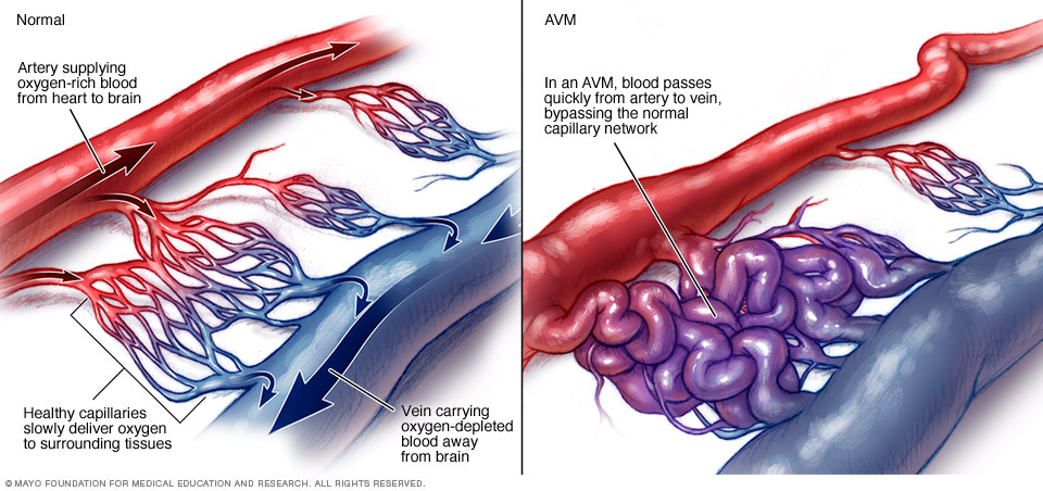
In an arteriovenous malformation (AVM), blood passes quickly from the artery to vein, disrupting the normal blood flow and depriving the surrounding tissues of oxygen.
The cause of brain AVM is unknown, but researchers believe most brain AVMs emerge during fetal development.
Normally, your heart sends oxygen-rich blood to your brain through arteries. The arteries slow blood flow by passing it through a series of progressively smaller networks of blood vessels, ending with the smallest blood vessels (capillaries). The capillaries slowly deliver oxygen through their thin, porous walls to the surrounding brain tissue.
The oxygen-depleted blood then passes into small blood vessels and then into larger veins that drain the blood from your brain, returning it to your heart and lungs to get more oxygen.
The arteries and veins in an AVM lack this supporting network of smaller blood vessels and capillaries. Instead, the abnormal connection causes blood to flow quickly and directly from your arteries to your veins, bypassing the surrounding tissues.
Risk factors
Anyone can be born with a brain AVM, but these factors may be a risk:
- Being male. AVMs are more common in males.
- Having a family history. Cases of AVMs in families have been reported, but it's unclear if there's a certain genetic factor or if the cases are only coincidental. It's also possible to inherit other medical conditions that predispose you to having vascular malformations such as AVMs.
Complications

A brain AVM may cause bleeding in the brain (hemorrhage), which can damage the surrounding brain tissue, as shown by this CT scan (left) and illustration (right) of an intracerebral hemorrhage.
Complications of a brain AVM include:
-
Bleeding in the brain (hemorrhage). An AVM puts extreme pressure on the walls of the affected arteries and veins, causing them to become thin or weak. This may result in the AVM rupturing and bleeding into the brain (a hemorrhage).
This risk of a brain AVM bleeding ranges around 2 percent each year. The risk of hemorrhage may be higher for certain types of AVMs, or if you have experienced previous AVM ruptures.
Some hemorrhages associated with AVMs go undetected because they cause no major brain damage or symptoms, but potentially life-threatening bleeding episodes may occur.
Brain AVMs account for about 2 percent of all hemorrhagic strokes each year and are often the cause of hemorrhage in children and young adults who experience brain hemorrhage.
-
Reduced oxygen to brain tissue. With an AVM, blood bypasses the network of capillaries and flows directly from arteries to veins. Blood rushes quickly through the altered path because it isn't slowed down by channels of smaller blood vessels.
Surrounding brain tissues can't easily absorb oxygen from the fast-flowing blood. Without enough oxygen, brain tissues weaken or may die off completely. This results in stroke-like symptoms, such as difficulty speaking, weakness, numbness, vision loss or severe unsteadiness.
- Thin or weak blood vessels. An AVM puts extreme pressure on the thin and weak walls of the blood vessels. A bulge in a blood vessel wall (aneurysm) may develop and become susceptible to rupture.
-
Brain damage. As you grow, your body may recruit more arteries to supply blood to the fast-flowing AVM. As a result, some AVMs may get bigger and displace or compress portions of the brain. This may prevent protective fluids from flowing freely around the hemispheres of the brain.
If fluid builds up, it can push brain tissue up against the skull (hydrocephalus).
Diagnosis
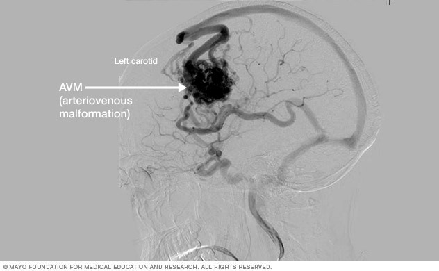
Cerebral angiogram showing brain AVM
To diagnose a brain AVM, your neurologist will review your symptoms and conduct a physical examination.
Your doctor may order one or more tests to diagnose your condition. Radiologists trained in brain and nervous system imaging (neuroradiologists) usually conduct imaging tests.
Tests used to diagnose brain AVMs include:
-
Cerebral arteriography. Cerebral arteriography, also known as cerebral angiography, is the most detailed test to diagnose an AVM. The test reveals the location and characteristics of the feeding arteries and draining veins, which is critical to planning treatment.
In this test, your doctor inserts a long, thin tube (catheter) into an artery in the groin and threads it to your brain using X-ray imaging. Your doctor injects dye into the blood vessels of your brain to make them visible under X-ray imaging.
-
Computerized tomography (CT) scan. A CT scan uses a series of X-rays to create a detailed cross-sectional image of your brain.
Sometimes a doctor injects dye through an intravenous tube into a vein so that the arteries feeding the AVM and the veins draining the AVM can be viewed in greater detail (computerized tomography angiogram).
-
Magnetic resonance imaging (MRI). MRI uses powerful magnets and radio waves to create detailed images of your brain.
MRI is more sensitive than CT and can show more subtle changes in brain tissue associated with a brain AVM.
MRI also provides information about the exact location of the malformation and any related bleeding in the brain, which is important for determining treatment options.
Your doctor may also inject dye to see the blood circulation in your brain (magnetic resonance angiogram).
Treatment
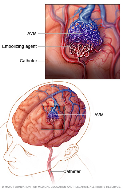
In endovascular embolization, your doctor inserts a long, thin tube (catheter) into a leg artery and threads it through blood vessels to your brain using X-ray imaging. Your surgeon positions the catheter in one of the feeding arteries to the AVM, and injects an embolizing agent, such as small particles or a glue-like substance, to block the artery and reduce blood flow into the AVM.
There are several potential treatment options for brain AVM. The main goal of treatment is to prevent hemorrhage, but treatment to control seizures or other neurological complications also may be considered.
Your doctor will determine the most appropriate treatment for your condition, depending on your age, health, and the size and location of the abnormal blood vessels.
Medications also may be used to treat symptoms caused by the AVM, such as headaches or seizures.
Surgery is the most common treatment for brain AVMs. There are three different surgical options for treating AVMs:
-
Surgical removal (resection). If the brain AVM has bled or is in an area that can easily be reached, surgical removal of the AVM via conventional brain surgery may be recommended. In this procedure, your neurosurgeon removes part of your skull temporarily to gain access to the AVM.
With the help of a high-powered microscope, the surgeon seals off the AVM with special clips and carefully removes it from surrounding brain tissue. The surgeon then reattaches the skull bone and closes the incision in your scalp.
Resection is usually done when the AVM can be removed with little risk of hemorrhage or seizures. AVMs that are in deep brain regions carry a higher risk of complications. In these cases, your doctor may recommend other treatments.
-
Endovascular embolization. In this procedure, your doctor inserts a long, thin tube (catheter) into a leg artery and threads it through blood vessels to your brain using X-ray imaging.
The catheter is positioned in one of the feeding arteries to the AVM, and injects an embolizing agent, such as small particles, a glue-like substance, microcoils or other materials, to block the artery and reduce blood flow into the AVM.
Endovascular embolization is less invasive than traditional surgery. It may be performed alone, but is frequently used prior to other surgical treatments to make the procedure safer by reducing the size of the AVM or the likelihood of bleeding.
In some large brain AVMs, endovascular embolization may be used to reduce stroke-like symptoms by redirecting blood back to normal brain tissue.
-
Stereotactic radiosurgery (SRS). This treatment uses precisely focused radiation to destroy the AVM. It is not surgery in the literal sense because there is no incision.
Instead, SRS directs many highly targeted radiation beams at the AVM to damage the blood vessels and cause scarring. The scarred AVM blood vessels then slowly clot off in one to three years following treatment.
This treatment is most appropriate for small AVMs that are difficult to remove with conventional surgery and for those that haven't caused a life-threatening hemorrhage.
If you have few or no symptoms or if your AVM is in an area of your brain that's hard to treat, your doctor may prefer to monitor your condition with regular checkups.
Potential future treatments
Researchers are currently studying ways to better predict the risk of hemorrhage in people with brain AVM to better guide treatment decisions. For example, high blood pressure within the AVM and hereditary syndromes associated with neurological issues may play a role.
Innovations in imaging technology, such as 3-D imaging, functional imaging and brain tract mapping also are being evaluated and have the potential to improve surgical precision and safety in removing brain AVMs and preserving surrounding vessels.
In addition, ongoing advances in embolization, radiosurgery and microsurgery techniques are making previously inoperable brain AVMs more accessible and safer for surgical removal.
Coping and support
Learning that you have a brain AVM can be frightening. It can make you feel like you have little control over your health. But you can take steps to cope with the emotions that accompany your diagnosis and recovery. Consider trying to:
- Learn enough about brain AVM to make informed decisions about your care. Ask your doctor about the size and location of your brain AVM and how that affects your treatment options. As you learn more about brain AVMs, you may become more confident in making treatment decisions.
- Accept your emotions. Complications of brain AVM, such as hemorrhage and stroke, can cause emotional problems as well as physical ones. Recognize that emotions may be hard to control, and some emotional and mood changes may be caused by the injury itself as well as coming to terms with the diagnosis.
- Keep friends and family close. Keeping your close relationships strong will help you during your recovery. Friends and family can provide the practical support you'll need, like accompanying you to doctors' appointments, and serve as emotional support.
- Find someone to talk with. Find a good listener who is willing to listen to you talk about your hopes and fears. This may be a friend or family member. The concern and understanding of a counselor, medical social worker, clergy member or support group also may be helpful.
Ask your doctor about support groups in your area. Or check your phone book, library or a national organization, such as the American Stroke Association or the Aneurysm and AVM Foundation.
Preparing for an appointment
A brain AVM may be diagnosed in an emergency situation, immediately after bleeding (hemorrhage) has occurred. It may also be detected after other symptoms prompt a brain scan.
But in some cases, a brain AVM is found during diagnosis or treatment of an unrelated medical condition. You may then be referred to a doctor trained in brain and nervous system conditions (neurologist or neurosurgeon).
Because there's often a lot to discuss, it's a good idea to arrive well-prepared for your appointment. Here are some tips to help you get ready for your appointment, and what to expect from your doctor.
What you can do
- Be aware of any pre-appointment restrictions. At the time you make the appointment, be sure to ask if there's anything you need to do in advance.
- Write down any symptoms you're experiencing, including any that may seem unrelated to the reason for which you scheduled the appointment.
- Make a list of all medications, vitamins and supplements that you're taking.
- Ask a family member or friend to come with you, if possible. Sometimes it can be difficult to absorb all the information provided to you during an appointment. Someone who accompanies you may remember something that you forgot or missed.
- Write down questions to ask your doctor. Don't be afraid to ask questions that may come up during your appointment.
Your time with your doctor is limited, so preparing a list of questions ahead of time will help you make the most of your time together. For brain AVM, some basic questions to ask your doctor include:
- What are other possible causes for my symptoms?
- What tests are needed to confirm the diagnosis?
- What are my treatment options and the pros and cons for each?
- What results can I expect?
- What kind of follow-up should I expect?
What to expect from your doctor
Your neurologist is likely to ask about your symptoms, if any, conduct a physical examination and schedule tests to confirm the diagnosis.
The tests gather information about the size and location of the AVM to help direct your treatment options. He or she may ask:
- When did you first begin experiencing symptoms?
- Have your symptoms been continuous or occasional?
- How severe are your symptoms?
- What, if anything, seems to improve your symptoms?
- What, if anything, appears to worsen your symptoms?
What you can do in the meantime
Avoid any activity that may raise your blood pressure and put strain on a brain AVM, such as heavy lifting or straining. Also avoid taking any blood-thinning medications, such as warfarin.
Content Last Updated: May 17, 2019
Content provided by Mayo Clinic ©1998-2026 Mayo Foundation for Medical Education and Research (MFMER). All rights reserved. Terms of Use