Ebstein anomaly
Overview
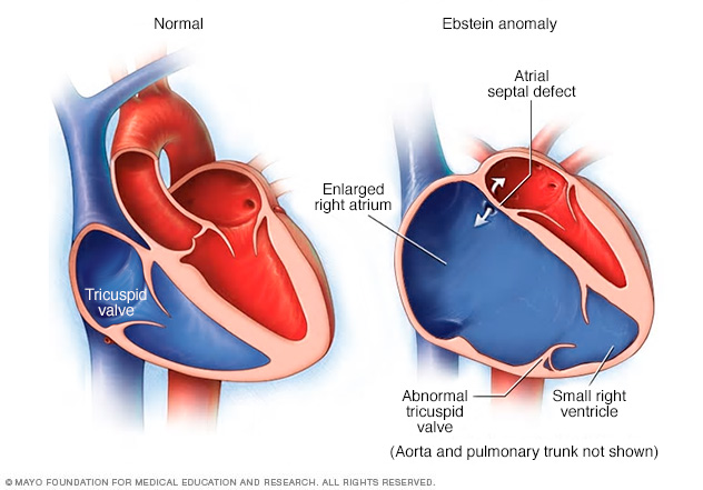
Ebstein anomaly is a rare congenital heart defect in which the tricuspid valve is incorrectly formed and positioned lower than usual in the heart. Atrial septal defect is a hole between the two upper chambers of the heart. About half the people with Ebstein anomaly have an atrial septal defect.
Ebstein anomaly is a rare heart defect that's present at birth (congenital). In this condition, your tricuspid valve is in the wrong position and the valve's flaps (leaflets) are malformed. As a result, the valve does not work properly.
Blood might leak back through the valve, making your heart work less efficiently. Ebstein anomaly can also lead to enlargement of the heart and heart failure.
If you have no signs or symptoms associated with Ebstein anomaly, regular monitoring of your heart might be all you need. You might need treatment if signs and symptoms bother you or if your heart is enlarging or weakening. Treatment options include medications and surgery.
Symptoms
Mild forms of Ebstein anomaly might not cause symptoms until later in adulthood. Signs and symptoms might include:
- Shortness of breath, especially with exertion
- Fatigue
- Heart palpitations or abnormal heart rhythms (arrhythmias)
- A bluish discoloration of the lips and skin caused by low oxygen (cyanosis)
When to see a doctor
If you or your child has signs or symptoms of heart failure — such as feeling easily fatigued or short of breath, even with normal activity — or if the skin around the lips and nails looks blue or you have swelling of your legs, talk to your doctor. He or she may refer you to a doctor who specializes in congenital heart disease (cardiologist).
Causes
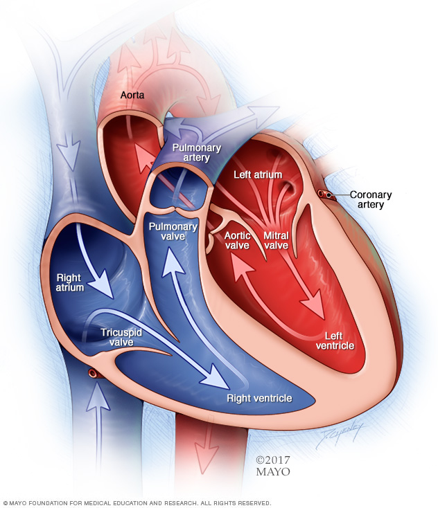
A typical heart has two upper and two lower chambers. The upper chambers — the right and left atria — receive incoming blood. The lower chambers — the right and left ventricles — pump blood out of the heart. The heart valves, which keep blood flowing in the right direction, are gates at the chamber openings (for the tricuspid and mitral valves) and exits (for the pulmonary and aortic valves).
Ebstein anomaly is a heart defect that you have at birth (congenital). The cause is unknown. To understand how Ebstein anomaly affects your heart, it helps to know how the heart works to supply your body with blood.
How your heart works
Your heart is made up of four chambers. The two upper chambers (atria) receive blood. The two lower chambers (ventricles) pump blood.
Four valves open and close to let blood flow in one direction through the heart. Each valve consists of two or three strong, thin flaps (leaflets) of tissue. A closed valve prevents blood from flowing to the next chamber or from returning to the previous chamber.
Oxygen-poor blood from your body flows into the right atrium. Blood then flows through the tricuspid valve into the right ventricle, which pumps the blood to your lungs. On the other side of your heart, oxygen-rich blood from your lungs flows into the left atrium, through the mitral valve and into the left ventricle, which then pumps the blood to the rest of your body.
What happens in Ebstein anomaly
The tricuspid valve normally sits between the two right heart chambers (right atrium and right ventricle).
In Ebstein anomaly, the tricuspid valve sits lower than normal in the right ventricle. This makes it so that a portion of the right ventricle becomes part of the right atrium, causing the right atrium to enlarge and not work properly.
Also, the tricuspid valve's leaflets are abnormally formed. This can lead to blood leaking backward into the right atrium (tricuspid valve regurgitation).
The location of the valve and how poorly it's formed varies from person to person. Some people have a mildly abnormal valve. Others have a valve that leaks severely.
Other heart conditions associated with Ebstein anomaly
Common associated heart conditions include:
-
Holes in the heart. Many people with Ebstein anomaly have a hole between the two upper chambers of the heart called an atrial septal defect or an opening called a patent foramen ovale (PFO). A PFO is a hole between the upper heart chambers that all babies have before birth that usually closes after birth. It can remain open in some people without causing issues.
These holes can decrease the amount of oxygen available in your blood, causing a bluish discoloration of the lips and skin (cyanosis).
- Abnormal heartbeats (arrhythmias). An abnormal heart rhythm or rapid heartbeats make it difficult for the heart to work properly, especially when the tricuspid valve is leaking severely. Sometimes, a very fast heart rhythm causes fainting spells (syncope).
- Wolff-Parkinson-White (WPW) syndrome. People with WPW syndrome have an abnormal electrical pathway in the heart that can lead to fast heart rates and fainting spells.
Risk factors
Congenital heart defects, such as Ebstein anomaly, occur as the baby's heart develops in the mother's womb.
Doctors aren't sure what risk factors are associated with the defect. Genetic and environmental factors are believed to play a role. A family history of heart defects or a mother's use of certain medications, such as lithium, during pregnancy might increase the risk of Ebstein anomaly in the child.
Complications
Mild Ebstein anomaly may not cause any complications.
However, possible complications of Ebstein anomaly include:
- Heart failure
- Sudden cardiac arrest
- Stroke
Taking a few precautions before playing sports or becoming pregnant may help prevent complications.
If your heart size is nearly normal and you have no heart rhythm disturbances, you can probably participate in most physical activities. Depending on your signs and symptoms, your doctor might recommend that you avoid certain competitive sports, such as football or basketball.
If you're planning on having a baby, talk to your doctor. Many women with mild Ebstein anomaly can safely have children. But pregnancy, labor and delivery put additional strain on your heart. Rarely, severe complications can develop that can cause death to mother or baby.
Together, you and your doctor can decide how much monitoring you need throughout pregnancy and childbirth. Sometimes, other treatments for your condition or symptoms may be recommended before you become pregnant.
Diagnosis
If you or your child doesn't have signs or symptoms of heart trouble, the doctor might suspect a problem only after hearing abnormal heart sounds during a routine physical exam.
Abnormal heart sounds, such as a heart murmur, aren't usually cause for concern. However, your doctor or your child's doctor will likely refer you to a doctor who specializes in treating heart conditions (cardiologist) to determine the cause.
Your doctor might recommend several tests, including:
-
Echocardiogram. This test uses sound waves to produce detailed images of your heart. It shows the structure of your tricuspid valve and the blood flow through your heart.
Sometimes, a transesophageal echocardiogram is done. This test uses a tube with a tiny sound device (transducer) inserted into the part of your digestive tract that runs from your throat to your stomach (esophagus). Because your esophagus lies close to your heart, this test can provide a detailed image of your heart.
- Electrocardiogram (ECG). Sensors (electrodes) attached to your chest and limbs measure the timing and duration of your heartbeat. An ECG can help show problems with your heart's rhythm and structure. Some personal devices, such as smartwatches, offer remote ECG monitoring. Ask your doctor if this is an option for you.
- Holter monitor. A Holter monitor is a portable ECG device that you wear while away from the doctor's office. It records your heart's electrical activity as you perform your normal activities for a day or two.
- Chest X-ray. A chest X-ray is a picture of your heart, lungs and blood vessels. It can tell your doctor if your heart is enlarged.
- Cardiac MRI. A cardiac MRI uses magnetic fields and radio waves to create detailed images of your heart. This test gives your doctor a detailed view of your tricuspid valve. It allows your doctor to see the size of your heart chambers and how well they work.
- Pulse oximetry. In this test, a sensor attached to your finger or toe measures the amount of oxygen in your blood.
- Exercise stress test. During this test, your blood pressure, heart rate, heart rhythm and breathing are monitored as you walk on a treadmill or ride a stationary bicycle. An exercise stress test can show how your heart responds to exercise. It can help your doctor decide what level of physical activity is safe for you.
-
Electrophysiology study (EP). To perform this test, the doctor threads thin, flexible tubes (catheters) tipped with electrodes through your blood vessels to areas within your heart to map your heart's electrical impulses.
In addition, your doctor can use the electrodes to stimulate your heart to beat at rates that may trigger — or halt — an arrhythmia. This can help your doctor determine if medications can help treat the arrhythmia.
- Cardiac catheterization. A long, thin tube (catheter) is inserted into a blood vessel in your groin, arm or neck and guided to your heart using X-ray imaging. A special dye injected through the catheter gives your doctor a clearer view of blood flow through your heart, blood vessels and valves. During the test, your doctor can measure pressures and oxygen levels in your heart and look for problems inside the heart and lungs.
Treatment
Treatment of Ebstein anomaly depends on the severity of the defect and your signs and symptoms. The goal of treatment is to reduce your symptoms and avoid future complications, such as heart failure and arrhythmias.
Regular monitoring
If you have no signs or symptoms or abnormal heart rhythms, your doctor might recommend monitoring your heart condition with regular checkups.
Follow-up appointments generally include a physical exam and tests such as an electrocardiogram, echocardiogram, Holter monitor test and exercise stress test.
Medications
If you have heart rhythm disturbances, medications might help control your heart rate and maintain normal heart rhythm.
Your doctor might also prescribe medications to ease signs and symptoms of heart failure, such as drugs to prevent water retention (diuretics).
If you have certain heart rhythm problems or a hole (atrial septal defect) between the upper heart chambers, your doctor may prescribe medications to prevent blood clots.
Some babies are given an inhaled substance called nitric oxide to help improve blood flow to the lungs.
Surgery or other procedures
Your doctor might recommend surgery if your symptoms are affecting your quality of life. Surgery might also be recommended if your heart is enlarging and your heart function is decreasing. If you do need surgery, it's important to choose a surgeon who's familiar with the defect and who has training and experience performing procedures to correct it.
Several types of procedures can be used to surgically treat Ebstein anomaly and associated defects.
-
Tricuspid valve repair. Surgeons reduce the size of the valve opening and allow the valve leaflets to come together to work properly. A band might be placed around the valve to keep it in place. This procedure is usually done when there's enough valve tissue to allow repair.
A newer form of tricuspid valve repair is called cone reconstruction. Surgeons separate the leaflets of the tricuspid valve from the heart muscle. The leaflets are then rotated and reattached, creating a "leaflet cone."
Sometimes, your valve might need to be repaired again or replaced in the future.
-
Tricuspid valve replacement. If the valve can't be repaired, your surgeon might remove it and replace it with either a biological tissue (bioprosthetic) or mechanical valve. Mechanical valves aren't used often for tricuspid valve replacement.
If you have a mechanical valve, you'll need a blood thinner to prevent blood clots. If you have any type of artificial valve, you'll need to take medication to prevent an inflammation of the inner lining of your heart (endocarditis) before dental procedures.
- Closure of the atrial septal defect. If there's a hole between the upper chambers of the heart (atrial septal defect), your surgeon can repair or replace the defective valve. Your surgeon can also repair other heart defects you have during this surgery.
-
Maze procedure. If you have fast heart rhythms, your surgeon may perform the Maze procedure during valve repair or replacement surgery. In this procedure, your surgeon makes small incisions in the upper chambers of your heart to create a pattern, or maze, of scar tissue.
Because scar tissue doesn't conduct electricity, it interrupts the stray heart signals that cause some types of arrhythmias. Extreme cold (cryotherapy) or heat (radiofrequency) energy also can be used to create the scars.
-
Radiofrequency catheter ablation. If you have fast or abnormal heart rhythms, your doctor might perform this procedure. Your doctor threads one or more catheters through your blood vessels to your heart.
Sensors at the tips of the catheters use heat (radiofrequency energy) to damage (ablate) a small area of heart tissue. This blocks the abnormal signals that are causing your arrhythmia. Some people may need repeat procedures.
- Heart transplantation. If you have severe Ebstein anomaly and poor heart function, a heart transplant might be necessary.
Coping and support
If you or your child has mild Ebstein anomaly, here's what you can do to manage symptoms and improve comfort.
- Follow up on medical care. See a cardiologist experienced in treating congenital heart disease for regular checkups. Report new or worsening signs or symptoms to the doctor. Timely treatment can keep the condition from worsening.
- Take medications as prescribed. Taking the right dose at the right time can help ease symptoms such as racing heartbeats, fatigue and shortness of breath.
- Stay active. Be as physically active as your or your child's doctor allows. Exercise can help strengthen the heart and improve blood flow. Encourage playtime with breaks as needed. Ask the doctor for a note you can give to your child's teachers or caregivers describing activity restrictions.
- Create a support network. Although many people with congenital heart defects lead normal, healthy lives, living with a heart defect can be challenging if you or your child needs specialized care. A serious health condition can create a physical, emotional and financial strain. Having family and friends you can rely on is critical. Some people find that support groups are a helpful source of information, comfort and friendship.
Preparing for an appointment
Your primary care doctor may refer you to a doctor who specializes in treating heart conditions (cardiologist). Here's some information to help you get ready for your appointment.
What you can do
When you make the doctor's appointment, be sure to ask if you need to do anything in advance, such as restrict your or your child's diet.
Write down the following information and take it with you to the appointment:
- Symptoms, including any that may seem unrelated to the reason for your appointment
- When the symptoms began
- All medications, vitamins and supplements currently being taken, including doses
- Questions to ask the doctor
If you're seeing a new doctor, request that a copy of medical records be sent to the new office.
For Ebstein anomaly, specific questions to ask your doctor include:
- What's the most likely cause of these symptoms?
- What kinds of tests do I or my child need?
- What treatments are available, and which do you recommend?
- What side effects can the treatment cause?
- How can I best manage this condition with other conditions I have or my child has?
- Do I or does my child need to restrict activity?
- Are there brochures or other printed material I can have? What websites do you recommend?
Don't hesitate to ask other questions.
What to expect from your doctor
Your doctor is likely to ask you questions, such as:
- How often do you have symptoms?
- Does anything seem to improve the symptoms?
- What, if anything, makes symptoms worse?
- What medications are you or your child taking?
Content Last Updated: June 18, 2021
Content provided by Mayo Clinic ©1998-2026 Mayo Foundation for Medical Education and Research (MFMER). All rights reserved. Terms of Use