Tetralogy of Fallot
Overview
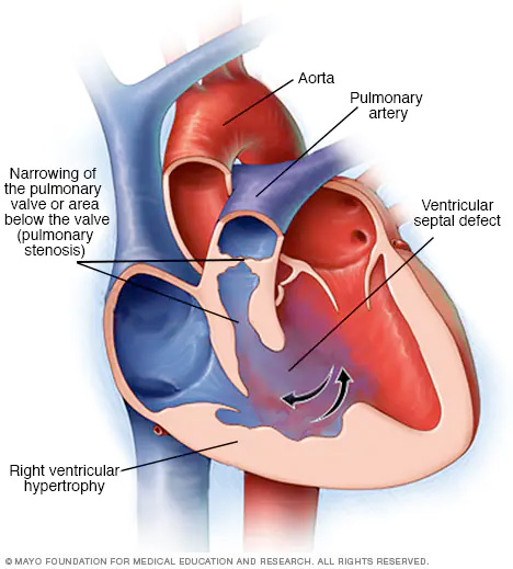
Tetralogy of Fallot is a combination of four congenital heart defects. The four defects are a ventricular septal defect (VSD), pulmonary stenosis, a misplaced aorta and a thickened right ventricular wall (right ventricular hypertrophy). They usually result in a lack of oxygen-rich blood reaching the body.
Tetralogy of Fallot (teh-TRAL-uh-jee of fuh-LOW) is a rare condition caused by a combination of four heart defects that are present at birth (congenital).
These defects, which affect the structure of the heart, cause oxygen-poor blood to flow out of the heart and to the rest of the body. Infants and children with tetralogy of Fallot usually have blue-tinged skin because their blood doesn't carry enough oxygen.
Tetralogy of Fallot is often diagnosed while the baby is an infant or soon after. Sometimes, depending on the severity of the defects and symptoms, tetralogy of Fallot is not detected until adulthood.
All babies who have tetralogy of Fallot need corrective surgery. People with tetralogy of Fallot need regular doctor's checkups for the rest of their life and may have activity restrictions.
Symptoms
Tetralogy of Fallot symptoms vary, depending on the amount of blood flow that's blocked. Signs and symptoms may include:
- A bluish coloration of the skin caused by low blood oxygen levels (cyanosis)
- Shortness of breath and rapid breathing, especially during feeding or exercise
- Poor weight gain
- Tiring easily during play or exercise
- Irritability
- Prolonged crying
- Heart murmur
- Fainting
- An abnormal, rounded shape of the nail bed in the fingers and toes (clubbing)
Tet spells
Sometimes, babies who have tetralogy of Fallot will suddenly develop deep blue skin, nails and lips after crying or feeding, or when agitated.
These episodes are called tet spells. Tet spells are caused by a rapid drop in the amount of oxygen in the blood. Tet spells are most common in young infants, around 2 to 4 months old. Toddlers or older children might instinctively squat when they're short of breath. Squatting increases blood flow to the lungs.
When to see a doctor
Seek medical help if you notice that your baby has the following signs or symptoms:
- Difficulty breathing
- Bluish discoloration of the skin
- Passing out or seizures
- Weakness
- Unusual irritability
If your baby becomes blue (cyanotic), place your baby on his or her side and pull your baby's knees up to his or her chest. This helps increase blood flow to the lungs. Call 911 or your local emergency number immediately.
Causes
Tetralogy of Fallot occurs as the baby's heart is developing during pregnancy. Usually, the cause is unknown.
Tetralogy of Fallot includes four defects:
- Narrowing of the lung valve (pulmonary valve stenosis). Narrowing of the valve that separates the lower right chamber of the heart (right ventricle) from the main blood vessel leading to the lungs (pulmonary artery) reduces blood flow to the lungs. The narrowing might also affect the muscle beneath the pulmonary valve. Sometimes, the pulmonary valve doesn't form properly (pulmonary atresia).
- A hole between the bottom heart chambers (ventricular septal defect). A ventricular septal defect is a hole in the wall (septum) that separates the two lower chambers of the heart (left and right ventricles). The hole causes oxygen-poor blood in the right ventricle to mix with oxygen-rich blood in the left ventricle. This causes inefficient blood flow and reduces the supply of oxygen-rich blood to the body. The defect eventually can weaken the heart.
- Shifting of the body's main artery (aorta). Normally the aorta branches off the left ventricle. In tetralogy of Fallot, the aorta is in the wrong position. It's shifted to the right and lies directly above the hole in the heart wall (ventricular septal defect). As a result, the aorta receives a mix of oxygen-rich and oxygen-poor blood from both the right and left ventricles.
- Thickening of the right lower heart chamber (right ventricular hypertrophy). When the heart's pumping action is overworked, the muscular wall of the right ventricle becomes thick. Over time this might cause the heart to stiffen, become weak and eventually fail.
Some children or adults who have tetralogy of Fallot may have other heart defects such as a hole between the heart's upper chambers (atrial septal defect), a right aortic arch or problems with the coronary arteries.
Risk factors
While the exact cause of tetralogy of Fallot is unknown, some things might increase the risk of a baby being born with this condition. Risk factors for tetralogy of Fallot include:
- A viral illness during pregnancy, such as rubella (German measles)
- Drinking alcohol during pregnancy
- Poor nutrition during pregnancy
- A mother older than age 40
- A parent who has tetralogy of Fallot
- The presence of Down syndrome or DiGeorge syndrome in the baby
Complications
A possible complication of tetralogy of Fallot is infection of the inner lining of the heart or heart valve caused by a bacterial infection (infective endocarditis). Your or your child's doctor may recommend taking antibiotics before certain dental procedures to prevent infections that might cause this infection.
People with untreated tetralogy of Fallot usually develop severe complications over time, which might result in death or disability by early adulthood.
Complications from tetralogy of Fallot surgery
While most babies and adults do well after open-heart surgery to repair tetralogy of Fallot defects (intracardiac repair), long-term complications are common. Complications may include:
- Leaking pulmonary valve (chronic pulmonary regurgitation), in which blood leaks through the valve back into the pumping chamber (right ventricle)
- Leaking tricuspid valve
- Holes in the wall between the ventricles (ventricular septal defects) that may continue to leak after repair or may need re-repair
- Enlarged right ventricle or left ventricle that isn't working properly
- Irregular heartbeats (arrhythmias)
- Coronary artery disease
- Enlargement of the ascending aorta (aortic root dilation)
- Sudden cardiac death
It's very important to have regular checkups with a heart doctor trained in caring for people with congenital heart disease (pediatric cardiologist or adult congenital cardiologist).
Diagnosis
Usually, tetralogy of Fallot is diagnosed soon after birth. Your baby's skin may appear blue. A doctor might hear an abnormal whooshing sound (heart murmur) when listening to the baby's heart with a stethoscope.
Tests to diagnose tetralogy of Fallot include:
- Oxygen level measurement (pulse oximetry). A small sensor placed on a finger or toe measures the amount of oxygen in the blood.
- Echocardiogram. An echocardiogram uses sound waves to create pictures of the heart in motion. An echocardiogram can show the structure, placement and function of the heart wall, heart chambers, heart and pulmonary valves, and aorta.
- Electrocardiogram (ECG or EKG). An electrocardiogram records the electrical activity in the heart each time it contracts. During this procedure, sticky patches with wires (electrodes) are placed on the chest, wrists and ankles. The wires connect to a computer, which displays the heart's rhythm. An ECG can help determine if the heart chambers are enlarged and if there's an abnormal heartbeat (arrhythmia).
- Chest X-ray. A chest X-ray can show the structure of the heart and lungs. A common sign of tetralogy of Fallot on an X-ray is a boot-shaped heart, because the right ventricle is enlarged.
-
Cardiac catheterization. Doctors may use this test to evaluate the structure of the heart and plan surgical treatment. During this procedure, the doctor inserts a thin, flexible tube (catheter) into a blood vessel, usually in the groin, and guides it to the heart.
Dye flows through the catheter to make the heart structures easier to see on X-rays. The doctor can measure pressure and oxygen levels in the heart's chambers and blood vessels during the procedure.
Treatment
All babies who have tetralogy of Fallot need corrective surgery performed by a heart (cardiovascular) surgeon. Without treatment, your baby might not grow and develop properly. Your doctor will determine the most appropriate surgery and the timing of the surgery based on your or your child's condition.
Some children may need medicine while waiting for surgery to maintain blood flow from the heart to the lungs.
Surgery or other procedures
Surgery for tetralogy of Fallot involves open-heart surgery to correct the defects (intracardiac repair) or a temporary procedure that uses a shunt. Most babies and older children have intracardiac repair.
Intracardiac repair
This open-heart surgery is usually done during the first year after birth and involves several repairs. Adults with tetralogy of Fallot rarely may undergo this procedure if they didn't have surgical repair as children.
During intracardiac repair, the surgeon will:
- Patch over the ventricular septal defect to close the hole between the lower chambers of the heart (ventricles).
- Repair or replace the narrowed pulmonary valve to increase blood flow to the lungs.
Because the right ventricle won't need to work as hard to pump blood after this procedure, the right ventricle wall will go back to its normal thickness. After intracardiac repair, the oxygen level in the blood increases and symptoms decrease.
Temporary shunt surgery
Occasionally babies need to undergo a temporary (palliative) surgery before having intracardiac repair in order to improve blood flow to the lungs. This procedure may be done if your baby was born prematurely or has pulmonary arteries that are undeveloped (hypoplastic).
In this procedure, the surgeon creates a bypass (shunt) between a large artery that branches off from the aorta and the pulmonary artery.
When your baby is ready for intracardiac repair, the surgeon removes the shunt during the procedure for intracardiac repair.
After surgery
The long-term survival rates for people who've had tetralogy of Fallot surgery continue to improve.
However, sometimes blood flow to the lungs may still be restricted after tetralogy of Fallot surgery. Additional surgeries may be needed. An adult with repaired tetralogy of Fallot may have a leaky pulmonary valve (pulmonary valve regurgitation) and may need to have their pulmonary valve eventually replaced.
Heart rhythm problems (arrhythmias) are common after tetralogy of Fallot repair surgery. Your doctor may recommend medications, a procedure to treat the arrhythmias (ablation) or a special pacemaker that treats life-threatening arrhythmias (implantable cardioverter-defibrillator).
Ongoing care
People with tetralogy of Fallot need lifelong care with a pediatric or adult congenital cardiologist to ensure the surgery's success and to monitor for complications. Checkups often include imaging tests to determine how well treatment is working.
Lifestyle and home remedies
After tetralogy of Fallot treatment, your doctor might recommend lifestyle changes and tips to help you manage your or your child's condition, including:
- Preventing infection. A child, adolescent or adult who has severe heart defects might need to take preventive antibiotics before certain dental procedures and surgeries. Your or your child's doctor can tell you if this is necessary. Maintaining good oral hygiene and getting regular dental checkups are important ways to help prevent infection.
- Limiting certain types of exercise. The doctor might recommend that you or your child limit strenuous physical activity, particularly if there are heart rhythm problems (arrhythmias) or leakage or blockage of the pulmonary valve. Decisions about exercise need to be made on an individual basis. Talk to your or your child's doctor about which activities are safe.
If you're an adult who has congenital heart disease, you might have other concerns, including:
- Employment. If you have serious heart rhythm problems or the potential for life-threatening complications, evaluation by a specialty team is needed to determine your specific risk, provide therapy and counsel you regarding employment.
- Pregnancy. A severe heart defect or arrhythmia can increase the risk of complications during pregnancy. If you have congenital heart disease, discuss family planning with your doctor. Your doctor may recommend that you receive care during your pregnancy from doctors trained in congenital heart disease, genetics and high-risk obstetrics. Some heart medications aren't safe during pregnancy and might need to be stopped or adjusted before you become pregnant.
Coping and support
It's natural to feel worried if you or your child are diagnosed with a congenital heart defect. Here are a few ways to help you ease stress and anxiety and best manage your or your child's condition.
- Join a support group. A support group allows you to share personal experiences and feelings with others who are going through similar challenges. Some people find that a support group gives them hope, encouragement and support. Ask your doctor if there are any support groups for parents of children with heart defects or adults with congenital heart disease in your area.
- Ask for help and take a break. If your child has a heart defect, be sure to give yourself a break at times. Ask other family members or friends to help take care of your child. When your child is in the hospital, see if you can schedule friends and family to visit with your child so that you can go home to take a shower or nap, or to spend time with your other children.
- Keep a diary. To help coordinate your or your child's care, you might keep a notebook with your or your child's diagnosis, medications, surgeries and dates, and the cardiologist's name and number. This information will be valuable to others who might care for your child and will help any new doctor understand your or your child's health history.
- Review your health insurance plan. If you change health insurance plans, be sure your new plan will cover your or your child's care. Some plans might not allow coverage for preexisting conditions or might require a waiting period.
Preparing for an appointment
You're likely to start by seeing your primary care doctor. You or your child will be referred to a doctor trained in treating heart conditions (cardiologist).
Here's some information to help you get ready for your appointment, and what to expect from your or your child's doctor.
What you can do
- Be aware of any pre-appointment restrictions. When you make the appointment, ask if there's anything you need to do in advance, such as restrict your or your child's diet.
- Write down any symptoms you or your baby is experiencing, including any that might seem unrelated to the reason for which you scheduled the appointment.
- Write down your or your child's family history, including details from both the maternal and paternal sides of the family.
- Ask a family member or friend to come with you, if possible. Sometimes it can be difficult to remember all of the information provided to you during an appointment.
- Write down questions to ask the doctor.
Preparing a list of questions can help you make the most of your appointment time. For tetralogy of Fallot, some basic questions to ask your or your child's doctor include:
- What's the most likely cause of my or my child's symptoms?
- Are there other possible causes of these symptoms?
- What kinds of tests do I or my child need? Do these tests require special preparation?
- What treatments are available, and which do you recommend?
- What are the possible complications of treatment?
- What's my or my child's outlook after surgery? Can I or my child live a normal life?
- My child or I have other health conditions. How can I best manage them together?
- Are there any activity restrictions?
- Will I or my child be able to play sports? Can my child participate in gym?
- Will this cause a problem during future pregnancies, and is there any way to prevent it?
- Are there any brochures or other printed material that I can take home with me? What websites do you recommend?
Don't hesitate to ask other questions during your appointment.
What to expect from your doctor
Your or your child's doctor is likely to ask you a number of questions, such as:
- When did you first notice symptoms?
- Do the symptoms occur all the time (continuous) or do they come and go (occasional)?
- Does anything seem to improve your or your child's symptoms?
- What, if anything, makes the symptoms worse?
- How are you or your child eating and sleeping?
- Have you noticed fainting spells or episodes when your or your child's lips and skin become more blue or dusky?
- Are you or your child vomiting or losing weight?
- Have you or your child had heart racing, breathlessness or leg swelling?
What you can do in the meantime
Here are a few tips to help make your child more comfortable:
- Feed your baby slowly. Try smaller, more frequent meals.
- Help your child during a tet spell. Your child's skin, nails and lips might turn blue after crying, feeding or waking up. If you can remain calm, it can help reduce your child's anxiety. Improve blood flow to your child's heart and lungs by gently raising his or her knees to the chest.
- In case of emergency, call 911 or your local emergency number or go to a hospital's emergency department.
Content Last Updated: August 17, 2021
Content provided by Mayo Clinic ©1998-2026 Mayo Foundation for Medical Education and Research (MFMER). All rights reserved. Terms of Use