Molar pregnancy
Overview
A molar pregnancy — also known as hydatidiform mole — is a rare complication of pregnancy characterized by the abnormal growth of trophoblasts, the cells that normally develop into the placenta.
There are two types of molar pregnancy, complete molar pregnancy and partial molar pregnancy. In a complete molar pregnancy, the placental tissue is abnormal and swollen and appears to form fluid-filled cysts. There's also no formation of fetal tissue. In a partial molar pregnancy, there may be normal placental tissue along with abnormally forming placental tissue. There may also be formation of a fetus, but the fetus is not able to survive, and is usually miscarried early in the pregnancy.
A molar pregnancy can have serious complications — including a rare form of cancer — and requires early treatment.
Symptoms
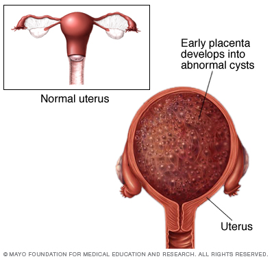
During a molar pregnancy, the placental tissue develops abnormally, and can appear as a mass of cysts. The embryo either doesn't form or is malformed and can't survive.
A molar pregnancy may seem like a normal pregnancy at first, but most molar pregnancies cause specific signs and symptoms, including:
- Dark brown to bright red vaginal bleeding during the first trimester
- Severe nausea and vomiting
- Sometimes vaginal passage of grapelike cysts
- Pelvic pressure or pain
If you experience any signs or symptoms of a molar pregnancy, consult your doctor or pregnancy care provider. He or she may detect other signs of a molar pregnancy, such as:
- Rapid uterine growth — the uterus is too large for the stage of pregnancy
- High blood pressure
- Preeclampsia — a condition that causes high blood pressure and protein in the urine after 20 weeks of pregnancy
- Ovarian cysts
- Anemia
- Overactive thyroid (hyperthyroidism)
Causes
A molar pregnancy is caused by an abnormally fertilized egg. Human cells normally contain 23 pairs of chromosomes. One chromosome in each pair comes from the father, the other from the mother.
In a complete molar pregnancy, an empty egg is fertilized by one or two sperm, and all of the genetic material is from the father. In this situation, the chromosomes from the mother's egg are lost or inactivated and the father's chromosomes are duplicated.
In a partial or incomplete molar pregnancy, the mother's chromosomes remain but the father provides two sets of chromosomes. As a result, the embryo has 69 chromosomes instead of 46. This most often occurs when two sperm fertilize an egg, resulting in an extra copy of the father's genetic material.
Risk factors
Approximately 1 in every 1,000 pregnancies is diagnosed as a molar pregnancy. Various factors are associated with molar pregnancy, including:
- Maternal age. A molar pregnancy is more likely in women older than age 35 or younger than age 20.
- Previous molar pregnancy. If you've had one molar pregnancy, you're more likely to have another. A repeat molar pregnancy happens, on average, in 1 out of every 100 women.
Complications
After a molar pregnancy has been removed, molar tissue may remain and continue to grow. This is called persistent gestational trophoblastic neoplasia (GTN). This occurs in about 15% to 20% of complete molar pregnancies, and up to 5% of partial molar pregnancies.
One sign of persistent GTN is a high level of human chorionic gonadotropin (HCG) — a pregnancy hormone — after the molar pregnancy has been removed. In some cases, an invasive hydatidiform mole penetrates deep into the middle layer of the uterine wall, which causes vaginal bleeding.
Persistent GTN can nearly always be successfully treated, most often with chemotherapy. Another treatment option is removal of the uterus (hysterectomy).
Rarely, a cancerous form of GTN known as choriocarcinoma develops and spreads to other organs. Choriocarcinoma is usually successfully treated with multiple cancer drugs. A complete molar pregnancy is more likely to have this complication than a partial molar pregnancy.
Prevention
If you've had a molar pregnancy, talk to your doctor or pregnancy care provider before conceiving again. He or she may recommend waiting for six months to one year before trying to become pregnant. The risk of recurrence is low, but higher than the risk for women with no previous history of molar pregnancy.
During any subsequent pregnancies, your care provider may do early ultrasounds to monitor your condition and offer reassurance of normal development. Your provider may also discuss prenatal genetic testing, which can be used to diagnose a molar pregnancy.
Diagnosis
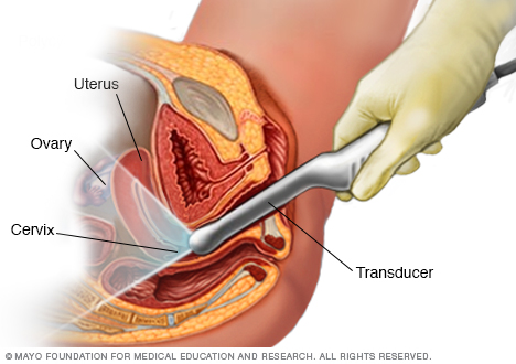
During a transvaginal ultrasound, your doctor or a medical technician inserts a wandlike device (transducer) into your vagina while you are positioned on an exam table. The transducer emits sound waves that generate images of your uterus, ovaries and fallopian tubes.
If your doctor suspects a molar pregnancy, he or she will order blood tests, including one to measure the level of human chorionic gonadotropin (HCG) — a pregnancy hormone — in your blood. He or she will also recommend an ultrasound.
With a standard ultrasound, high-frequency sound waves are directed at the tissues in the abdominal and pelvic area. During early pregnancy, however, the uterus and fallopian tubes are closer to the vagina than to the abdominal surface, so the ultrasound may be done through a wandlike device placed in your vagina.
An ultrasound of a complete molar pregnancy — which can be detected as early as eight or nine weeks of pregnancy — may show:
- No embryo or fetus
- No amniotic fluid
- A thick cystic placenta nearly filling the uterus
- Ovarian cysts
An ultrasound of a partial molar pregnancy may show:
- A fetus that's unexpectedly small for gestational age
- Low amniotic fluid
- Placenta that appears abnormal
If your health care provider detects a molar pregnancy, he or she may check for other medical problems, including:
- Preeclampsia
- Hyperthyroidism
- Anemia
Treatment
A molar pregnancy can't continue as a normal viable pregnancy. To prevent complications, the abnormal placental tissue must be removed. Treatment usually consists of one or more of the following steps:
-
Dilation and curettage (D&C). To treat a molar pregnancy, your doctor will remove the molar tissue from your uterus with a procedure called dilation and curettage (D&C). A D&C is usually done as an outpatient procedure in a hospital.
During the procedure, you'll receive a local or general anesthetic and be positioned on the operating room table on your back with your legs in stirrups. Your doctor will insert a speculum into your vagina, as in a pelvic exam, to see your cervix. He or she will then dilate your cervix and remove uterine tissue with a vacuum device.
- Hysterectomy. Rarely, if there is increased risk of gestational trophoblastic neoplasia (GTN) and there's no desire for future pregnancies, the uterus may be removed (hysterectomy).
-
HCG monitoring. After the molar tissue is removed, your doctor will repeat measurements of your HCG level until it returns to normal. If you continue to have HCG in your blood, you may need additional treatment.
Once treatment for the molar pregnancy is complete, your doctor may continue to monitor your HCG levels for six months to one year to make sure there's no remaining molar tissue.
Because pregnancy HCG levels also increase during a normal pregnancy, your doctor may recommend you wait six to 12 months before trying to become pregnant again. Your provider will recommend a reliable form of birth control during this time.
Coping and support
Losing a pregnancy is devastating. Give yourself time to grieve. Talk about your feelings and allow yourself to experience them fully. Turn to your partner, family and friends for support. If you're having trouble handling your emotions, consult your pregnancy care provider or a counselor.
Preparing for an appointment
You're likely to start by talking with your family doctor or pregnancy care provider. Here's some information to help you get ready for your appointment and know what to expect from your doctor.
What you can do
Before your appointment:
- Write down any symptoms you're experiencing, including when they first started and how they've changed over time.
- Make a note of the date of your last menstrual period, if you remember it.
- Write down key personal information, including any other medical conditions for which you're being treated.
- Make a list of all medications, as well as any vitamins or supplements you're taking.
- Ask a friend or family member to accompany you, if possible, to your appointment. Having someone else there may help you remember something that you forgot or missed and may provide much-needed emotional support.
- Write down questions to ask your doctor.
Preparing a list of questions in advance will help you make the most of your time with your doctor. For molar pregnancy, some basic questions to ask include:
- What is likely causing my symptoms or condition?
- What kind of tests do I need?
- What needs to be done now?
- What treatment approach do you recommend?
- Do I need to follow any restrictions?
- What emergency signs and symptoms should I watch for at home?
- What are my chances for a successful future pregnancy?
- How long should I wait before trying to become pregnant again?
- Does my condition increase my risk of developing cancer in the future?
- Do you have any brochures or printed material that I can take with me? What websites do you recommend for more information?
In addition to the questions that you've prepared to ask your doctor, don't hesitate to ask questions during your appointment if you don't understand something.
What to expect from your doctor
Your doctor will likely perform a physical exam and run some tests, including a blood test and ultrasound exam. He or she may also ask you a number of questions, such as:
- When was your last menstrual period?
- When did you first begin experiencing symptoms?
- Have your symptoms been continuous or occasional?
- Are you having any pain?
- Compared with your heaviest days of menstrual flow, is your bleeding more, less or about the same? Have you passed any grapelike cysts from your vagina?
- Are you experiencing any lightheadedness or dizziness?
- Have you had a past molar pregnancy?
- What chronic conditions, if any, do you have?
- Do you wish to become pregnant in the future?
Content Last Updated: May 13, 2021
Content provided by Mayo Clinic ©1998-2026 Mayo Foundation for Medical Education and Research (MFMER). All rights reserved. Terms of Use