Chest X-rays
Overview
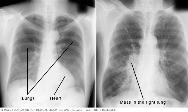
A chest X-ray helps detect problems with your heart and lungs. The chest X-ray on the left is normal. The image on the right shows a mass in the right lung.
Chest X-rays produce images of your heart, lungs, blood vessels, airways, and the bones of your chest and spine. Chest X-rays can also reveal fluid in or around your lungs or air surrounding a lung.
If you go to your doctor or the emergency room with chest pain, a chest injury or shortness of breath, you will typically get a chest X-ray. The image helps your doctor determine whether you have heart problems, a collapsed lung, pneumonia, broken ribs, emphysema, cancer or any of several other conditions.
Some people have a series of chest X-rays done over time to track whether a health problem is getting better or worse.
Why it's done
Chest X-rays are a common type of exam. A chest X-ray is often among the first procedures you'll have if your doctor suspects heart or lung disease. A chest X-ray can also be used to check how you are responding to treatment.
A chest X-ray can reveal many things inside your body, including:
- The condition of your lungs. Chest X-rays can detect cancer, infection or air collecting in the space around a lung, which can cause the lung to collapse. They can also show chronic lung conditions, such as emphysema or cystic fibrosis, as well as complications related to these conditions.
- Heart-related lung problems. Chest X-rays can show changes or problems in your lungs that stem from heart problems. For instance, fluid in your lungs can be a result of congestive heart failure.
- The size and outline of your heart. Changes in the size and shape of your heart may indicate heart failure, fluid around the heart or heart valve problems.
- Blood vessels. Because the outlines of the large vessels near your heart — the aorta and pulmonary arteries and veins — are visible on X-rays, they may reveal aortic aneurysms, other blood vessel problems or congenital heart disease.
- Calcium deposits. Chest X-rays can detect the presence of calcium in your heart or blood vessels. Its presence may indicate fats and other substances in your vessels, damage to your heart valves, coronary arteries, heart muscle or the protective sac that surrounds the heart. Calcified nodules in your lungs are most often from an old, resolved infection.
- Fractures. Rib or spine fractures or other problems with bone may be seen on a chest X-ray.
- Postoperative changes. Chest X-rays are useful for monitoring your recovery after you've had surgery in your chest, such as on your heart, lungs or esophagus. Your doctor can look at any lines or tubes that were placed during surgery to check for air leaks and areas of fluid or air buildup.
- A pacemaker, defibrillator or catheter. Pacemakers and defibrillators have wires attached to your heart to help control your heart rate and rhythm. Catheters are small tubes used to deliver medications or for dialysis. A chest X-ray usually is taken after placement of such medical devices to make sure everything is positioned correctly.
Risks
You may be concerned about radiation exposure from chest X-rays, especially if you have them regularly. But the amount of radiation from a chest X-ray is low — even lower than what you're exposed to through natural sources of radiation in the environment.
Even though the benefits of an X-ray outweigh the risk, you may be given a protective apron if you need multiple images. Tell your doctor if you're pregnant or might be pregnant. The procedure can be performed in a way to protect your abdomen from the radiation.
How you prepare
Before the chest X-ray, you generally undress from the waist up and wear an exam gown. You'll need to remove jewelry from the waist up, too, since both clothing and jewelry can obscure the X-ray images.
What you can expect
During the procedure, your body is positioned between a machine that produces the X-rays and a plate that creates the image digitally or with X-ray film. You may be asked to move into different positions in order to take views from both the front and the side of your chest.
During the front view, you stand against the plate, hold your arms up or to the sides and roll your shoulders forward. The X-ray technician may ask you to take a deep breath and hold it for several seconds. Holding your breath after inhaling helps your heart and lungs show up more clearly on the image.
During the side views, you turn and place one shoulder on the plate and raise your hands over your head. Again, you may be asked to take a deep breath and hold it.
Having X-rays taken is generally painless. You don't feel any sensation as the radiation passes through your body. If you have trouble standing, you may be able to have the exam while seated or lying down.
Results
A chest X-ray produces a black-and-white image that shows the organs in your chest. Structures that block radiation appear white, and structures that let radiation through appear black.
Your bones appear white because they are very dense. Your heart also appears as a lighter area. Your lungs are filled with air and block very little radiation, so they appear as darker areas on the images.
A radiologist — a doctor trained to interpret X-rays and other imaging exams — analyzes the images, looking for clues that may suggest if you have heart failure, fluid around your heart, cancer, pneumonia or another condition.
Your own doctor will discuss the results with you as well as what treatments or other tests or procedures may be necessary.
Content Last Updated: May 2, 2020
Content provided by Mayo Clinic ©1998-2026 Mayo Foundation for Medical Education and Research (MFMER). All rights reserved. Terms of Use