X-ray
Overview
An X-ray is a quick, painless test that produces images of the structures inside your body — particularly your bones.
X-ray beams pass through your body, and they are absorbed in different amounts depending on the density of the material they pass through. Dense materials, such as bone and metal, show up as white on X-rays. The air in your lungs shows up as black. Fat and muscle appear as shades of gray.
For some types of X-ray tests, a contrast medium — such as iodine or barium — is introduced into your body to provide greater detail on the images.
Why it’s done
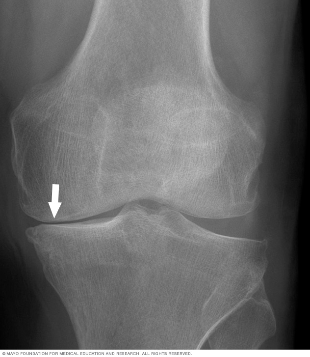
Knee arthritis can affect one side of the joint more than the other. This X-ray image shows how the cushioning cartilage has worn away, allowing bone to touch bone.
X-ray technology is used to examine many parts of the body.
Bones and teeth
- Fractures and infections. In most cases, fractures and infections in bones and teeth show up clearly on X-rays.
- Arthritis. X-rays of your joints can reveal evidence of arthritis. X-rays taken over the years can help your doctor determine if your arthritis is worsening.
- Dental decay. Dentists use X-rays to check for cavities in your teeth.
- Osteoporosis. Special types of X-ray tests can measure your bone density.
- Bone cancer. X-rays can reveal bone tumors.
Chest
- Lung infections or conditions. Evidence of pneumonia, tuberculosis or lung cancer can show up on chest X-rays.
- Breast cancer. Mammography is a special type of X-ray test used to examine breast tissue.
- Enlarged heart. This sign of congestive heart failure shows up clearly on X-rays.
- Blocked blood vessels. Injecting a contrast material that contains iodine can help highlight sections of your circulatory system to make them visible on X-rays.
Abdomen
- Digestive tract problems. Barium, a contrast medium delivered in a drink or an enema, can help reveal problems in your digestive system.
- Swallowed items. If your child has swallowed something such as a key or a coin, an X-ray can show the location of that object.
Risks
Radiation exposure
Some people worry that X-rays aren’t safe because radiation exposure can cause cell mutations that may lead to cancer. The amount of radiation you’re exposed to during an X-ray depends on the tissue or organ being examined. Sensitivity to the radiation depends on your age, with children being more sensitive than adults.
Generally, however, radiation exposure from an X-ray is low, and the benefits from these tests far outweigh the risks.
However, if you are pregnant or suspect that you may be pregnant, tell your doctor before having an X-ray. Though the risk of most diagnostic X-rays to an unborn baby is small, your doctor may consider another imaging test, such as ultrasound.
Contrast medium
In some people, the injection of a contrast medium can cause side effects such as:
- A feeling of warmth or flushing
- A metallic taste
- Lightheadedness
- Nausea
- Itching
- Hives
Rarely, severe reactions to a contract medium occur, including:
- Severe low blood pressure
- Anaphylactic shock
- Cardiac arrest
How you prepare
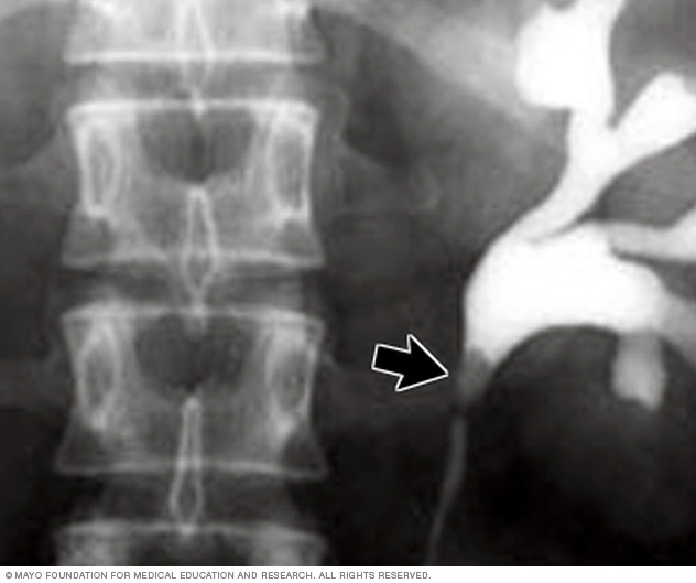
This X-ray using contrast reveals a kidney stone at the junction of the kidney and the tube that connects the kidney to the bladder (ureter).
Different types of X-rays require different preparations. Ask your doctor or nurse to provide you with specific instructions.
What to wear
In general, you undress whatever part of your body needs examination. You may wear a gown during the exam, depending on which area is being X-rayed. You may also be asked to remove jewelry, eyeglasses and any metal objects because they can show up on an X-ray.
Contrast material
Before some types of X-rays, you’re given a liquid called contrast medium. Contrast mediums, such as barium and iodine, help outline a specific area of your body on the X-ray image. You may swallow the contrast medium or receive it as an injection or an enema.
What you can expect
During the X-ray
X-rays are performed at doctors’ offices, dentists’ offices, emergency rooms and hospitals — wherever an X-ray machine is available. The machine produces a safe level of radiation that passes through your body and records an image on a specialized plate. You can’t feel an X-ray.
A technologist positions your body to obtain the necessary views. He or she may use pillows or sandbags to help you hold the position. During the X-ray exposure, you remain still and sometimes hold your breath to avoid moving so that the image doesn’t blur.
An X-ray procedure may take just a few minutes for a simple X-ray or longer for more-involved procedures, such as those using a contrast medium.
Your child’s X-ray
If a young child is having an X-ray, restraints or other techniques may be used to keep him or her still. These won’t harm your child and will prevent the need for a repeat procedure, which may be necessary if the child moves during the X-ray exposure.
You may be allowed to remain with your child during the test. If you remain in the room during the X-ray exposure, you’ll likely be asked to wear a lead apron to shield you from unnecessary exposure.
After the X-ray
After an X-ray, you generally can resume normal activities. Routine X-rays usually have no side effects. However, if you’re injected with contrast medium before your X-rays, drink plenty of fluids to help rid your body of it. Call your doctor if you have pain, swelling or redness at the injection site. Ask your doctor about other signs and symptoms to watch for.
Results
X-rays are saved digitally on computers, which can be viewed on-screen within minutes. A radiologist typically views and interprets the results and sends a report to your doctor, who then explains the results to you. In an emergency, your X-ray results can be made available to your doctor in minutes.
Content Last Updated: February 5, 2020
Content provided by Mayo Clinic ©1998-2022 Mayo Foundation for Medical Education and Research (MFMER). All rights reserved. Terms of Use